ADVANCED DENTAL IMAGING
See More, Plan Better, Treat with Confidence
Every diagnosis, every treatment plan starts with clear, detailed imaging. From digital x-rays to 3D CBCT scans and digital impressions, our technology reveals what's needed for precise, predictable care.
Advanced imaging means fewer surprises, more accurate treatment, and results that feel effortless—all available in-house.
DIGITAL X-RAYS
Safe, Fast, and Crystal Clear
Modern digital x-rays use 80-90% less radiation than traditional film while producing sharper images. The sensor captures instantly—no waiting, no chemical processing.
Each operatory has sensors and displays, so we review images together in real-time. Digital technology also means easy sharing with specialists or insurance, and secure storage in our system.
Types of X-Rays We Use
- •Bitewing X-Rays – Upper and lower teeth in one view, ideal for detecting cavities between teeth
- •Periapical X-Rays – Full tooth from crown to root tip, for diagnosing infections or abscesses
- •Full Mouth Series – Complete set showing all teeth and surrounding bone
When X-Rays Are Needed
New patients typically need a full set to establish a baseline. After that, most need bitewings every 1-2 years. We recommend based on individual risk factors—not a fixed schedule.
If you're experiencing pain, sensitivity, or swelling, diagnostic x-rays help us pinpoint the problem quickly.
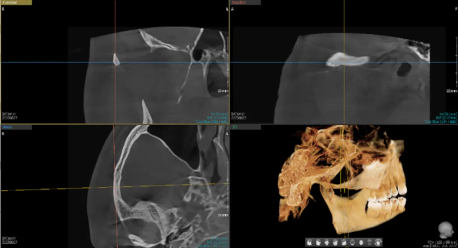
CBCT 3D IMAGING
Three-Dimensional Precision for Complex Cases
CBCT (Cone Beam Computed Tomography) takes dental imaging to another level. Instead of flat, two-dimensional images, CBCT creates a complete 3D reconstruction of your teeth, jaw bone, nerves, sinuses, and TMJ joints—all in a single scan.
Think of it as GPS for dentistry. We can rotate the image, measure bone density, identify nerve pathways, and plan treatments with millimeter precision. Most dental offices don't have this technology in-house, which means patients often need separate appointments at imaging centers. We bring that capability to you.
2D vs. 3D: The difference in diagnostic detail
When We Use CBCT Imaging
Dental Implant Planning
See exact bone height, width, and density. Identify nerve locations. Plan implant angle and depth with precision—reducing surgical time and improving outcomes.
Impacted Wisdom Teeth Evaluation
Visualize tooth position in 3D. Assess proximity to nerves and sinuses. Determine the safest extraction approach before surgery.
Root Canal Assessment
Detect extra canals or unusual anatomy that 2D x-rays miss. Locate fractures or infections. Plan endodontic treatment with confidence.
TMJ & Jaw Joint Analysis
Evaluate joint structure and movement. Assess bone changes or arthritis. Guide treatment for jaw pain or dysfunction.
Orthodontic Planning
Measure airway dimensions. Assess impacted teeth. Plan tooth movement with complete anatomical context.
The scanning process takes about 20-40 seconds. You'll stand or sit still while the scanner rotates once around your head. The radiation dose is a fraction of a medical CT scan—roughly equivalent to a few days of natural background radiation.
Images are available immediately. We'll review them with you on screen, pointing out areas of interest and explaining what we're seeing. This clarity helps you understand your diagnosis and feel confident about recommended treatment.
"Having CBCT in-house means you get answers in one visit. No separate appointments at imaging centers, no waiting for results to be sent over—just clear information when you need it."
Panoramic X-Rays: The Complete Picture
Panoramic x-rays capture your entire mouth—all teeth, upper and lower jaws, sinuses, and TMJ joints—in a single, wide-angle image. It's like a dental "panorama" that shows relationships between structures.
We use panoramic x-rays for comprehensive exams, orthodontic planning, wisdom teeth evaluation, and screening for jaw abnormalities. The process takes about 10 seconds—you simply stand still while the machine rotates around your head.
See What We See: Intraoral Cameras
A picture is worth a thousand words. Our intraoral cameras are tiny, pen-sized devices that capture high-resolution images of individual teeth. We can show you exactly what we're seeing—a cracked filling, early cavity, worn enamel, or gum recession.
This technology turns diagnosis into a conversation. When you see what we see, treatment decisions feel more informed and collaborative. No more wondering "do I really need that filling?"—you can see the cavity yourself.
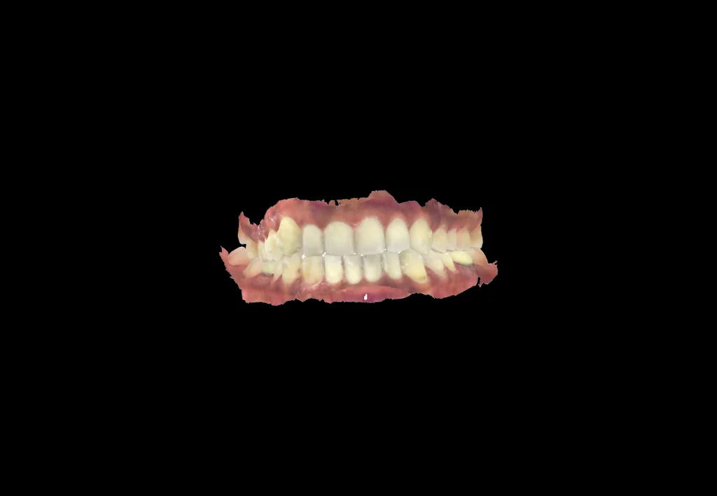
DIGITAL IMPRESSIONS
No More Gagging
Traditional impressions meant biting down on goopy trays and waiting uncomfortably. Digital impressions eliminate that entirely.
Our iTero and 3Shape scanners capture a precise 3D model in seconds. A small wand moves around your mouth—no gagging, no bad taste, no waiting for material to set. The digital process is faster, more comfortable, and more accurate.
We use digital impressions for Invisalign, crowns, bridges, veneers, and implant restorations. Better precision means better-fitting results with fewer adjustments.
The iTero Advantage for Invisalign
Considering Invisalign? The iTero scanner shows you a simulation of your future smile immediately—before you commit to treatment. Seeing the potential outcome helps you move forward with confidence.
Advanced Technology, Minimal Radiation
Radiation safety is one of the most common concerns about dental x-rays. Modern digital imaging has changed the equation dramatically.
| Scan Type | Dose (mSv) | Equivalent To... |
|---|---|---|
| Digital Bitewing X-ray | ~0.005 mSv | ~1 day of background radiation |
| Panoramic X-ray | ~0.025 mSv | ~3 days of background radiation |
| CBCT Scan (small field) | ~0.18 mSv | ~22 days of background radiation |
| Medical Head CT | ~2.0 mSv | ~8 months of background radiation |
| Annual Background Radiation (US average) | ~3.0 mSv | Natural yearly exposure from sun, soil, cosmic rays |
For perspective: A cross-country flight exposes you to about 0.035 mSv of radiation—more than a panoramic x-ray. Eating a banana gives you about 0.0001 mSv due to naturally occurring potassium-40.
Our digital x-rays use 80-90% less radiation than traditional film, and CBCT scans use approximately 10-20 times less radiation than medical CT scans while providing the detail we need for dental diagnosis.
We follow strict safety protocols:
- •Lead aprons and thyroid collars during x-rays
- •High-speed sensors that require minimal exposure time
- •Selective imaging—we only take x-rays when diagnostically necessary
- •Digital technology that eliminates retakes due to poor image quality
- •Customized settings for children and adults following ALARA principles (As Low As Reasonably Achievable)
If you're pregnant or might be pregnant, always let us know. We can often postpone elective x-rays until after delivery, or use additional protective measures if imaging is medically necessary.
References
[1] Radiological Society of North America (RSNA) – Radiation Dose in X-Ray and CT Exams. radiologyinfo.org/safety-xray
[2] Pereira et al., 2024 – Cone-Beam Computed Tomography Doses. American Academy of Pediatrics Journal. aap.onlinelibrary.wiley.com
[3] U.S. Environmental Protection Agency – Radiation Protection. epa.gov/radiation
Common Questions About Dental Imaging
Advanced Imaging Supports Every Treatment
From routine checkups to complex smile reconstruction, diagnostic imaging guides every decision we make. Clear information leads to confident treatment and predictable results.
See how we use imaging for: